

GALLBLADDER AND BILE DUCTS
Gallstones
Overview
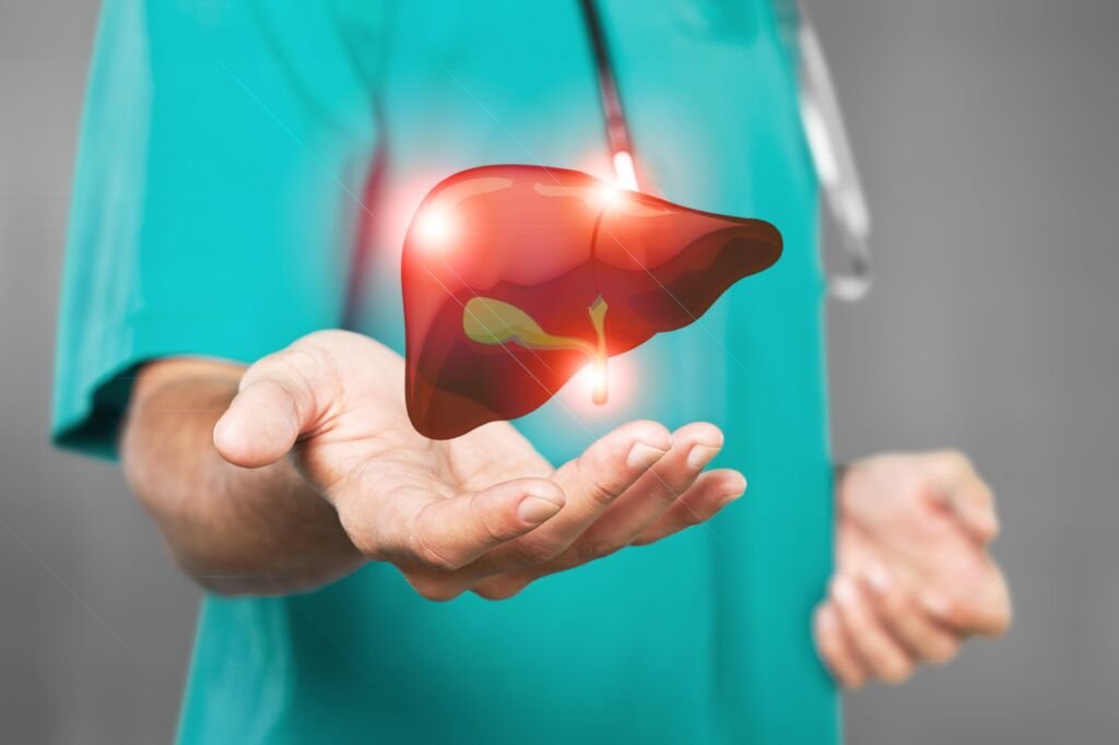
The gallbladder is a pear-shaped organ located beneath the liver at the right upper quadrant of the abdomen. It stores and concentrates bile which is secreted from the liver throughout the day. It contracts and empties the concentrated bile only when we eat. It contains a lot of excretory materials like cholesterol and bilirubin (Hemoglobin degradation by-product).
When the bile is oversaturated with products like cholesterol or bilirubin, they get hardened and form gallstones. Other than this, if the contraction of the gallbladder is not proper, it can also lead to stasis and stone formation.
Size of the stone:
The stone size varies from sand-like material (sludge) to lemon size. Gallstone may be single or multiple. The size and number of stones normally don’t matter to decide the treatment. It’s only the symptom results from the stone matters.
Types of gallstones:
- Cholesterol gallstones- yellow in color
- Pigment gallstones – contains bilirubin. Black/brown in color
- Mixed type – combination of cholesterol/bilirubin/other materials
Causes:
It’s difficult to pinpoint exactly what causes gallstone in a particular patient. It may be due to
- Too much cholesterol in bile.Cholesterol is normally excreted through the liver into bile. Bile also contains some natural chemicals (Lecithin) to dissolve cholesterol. It there is an imbalance (excess cholesterol or lack of dissolving chemicals), it may lead to crystallization of cholesterol which will eventually form stones.
- Too much bilirubin. Bilirubin is a breakdown product of hemoglobin in red blood cells. If bile contains too much bilirubin like in hemolytic disorders (red cell destruction), this will lead to stone formation
- Gallbladder contractile dysfunction: If the contractile function is not proper, this will leads to stasis of bile inside the gallbladder and encourage stone formation. E.g. Pregnancy

Symptoms:
Gallstones may or may not be symptomatic. It may cause a variety of symptoms depending on the location of the block it causes in the biliary tract. If a gallstone blocks the neck/cystic duct temporarily, it causes pain alone. It usually short-lasting and felt in the epigastric/right upper quadrant of the abdomen. In medical terminology, it is called biliary colic.

Acute calculous cholecystitis:
If a gallstone blocks the neck/cyst duct permanently, this will lead to stasis bile inside gallbladder and infection. This condition is called acute calculous cholecystitis. Sometimes if the infection is severe, the gallbladder will be filled with pus (Empyema gallbladder) or gangrene/perforation of the gallbladder. When this occurs, the patient will develop severe pain in the upper abdomen. This pain can radiate to the right shoulder/back. There may be associated fever/vomiting. Unlike biliary colic, this will be a continuous pain that often needs injectable analgesics (pain killers) or hospital admission.

Choledocholithiasis:
If gallstones enter into the common bile duct, it will be called choledocholithiasis. This may block the bile flow and cause jaundice, pain, and fever. Liver function tests will show abnormally elevated bilirubin and enzymes level. Obstruction of bile flow along with superadded infection can lead to a life-threatening infection called cholangitis. In this condition, the bacteria, as well as toxins from the biliary tree, enter into blood circulation. If the block has not been relieved promptly, the patient may go into septic shock and multiorgan failure.

Acute pancreatitis:
In some patients, the gallstones slip into the common bile duct and block the common channel of bile and pancreatic duct (Ampulla). Again, this is one of the dreaded complications of gallstones. Acute pancreatitis may be mild or severe types. Patients usually have upper abdominal pain often radiating to back and vomiting. In this condition, scans may not show any pancreatic abnormality in the initial few days. But the pancreatic enzymes like amylase/lipase will be elevated. Only in severe type of pancreatitis, patients go into multi-organ failure and often developed necrosis of pancreas (dead pancreatic tissue) with collections

Asymptomatic Gallstones:
Gallstones not associated with symptoms have been observed over a long time successfully. Only a small group of people developed problems, mostly pain to start with. The concept of observing (without surgery) asymptomatic gall stones is becoming common nowadays. But there are some medical exceptions to do surgery even if the stones are asymptomatic like hemolytic disorders.

Gallbladder cancer:
Although it is not proven beyond doubt, there is a suspicion that gallstones may be associated with gallbladder cancer. More or less, the likelihood of gallbladder cancer is very less

Gallstone ileus:
Rarely large gallstones perforate the intestine directly (bilio-enteric fistula) and block the intestine. Patients present with acute abdominal distension, pain and vomiting.

Risk factors for gallstone development:
- Being female
- Being age 40 or older
- Being overweight or obese
- Being pregnant
- Having a family history of gallstones
- Losing weight very quickly
- oral contraceptives or hormone therapy drugs
- Having chronic liver disease
Diagnosis
Blood test
The liver function test will be normal in asymptomatic/ uncomplicated gallstones. Elevated bilirubin/Alkaline phosphatase along with other enzyme abnormalities indicates complicated gallstone. Total leukocyte counts may be elevated in the presence of infection. Amylase/lipase enzymes are elevated in acute pancreatitis.
Imaging
Ultrasonography: Routine first investigation of gallstone disease is ultrasonography. It will pick up gallstones in most of the cases. However, if we suspect stones in the common bile duct, we need to do further imaging like MRCP (Magnetic resonance cholangiopancreatography)
MRCP
This is a form of Magnetic resonance imaging (MRI) which gives a map of the biliary and pancreatic ducts. It will show stones /block in the common bile duct.
ERCP
This is an invasive test usually done by endoscopists under anesthesia. Through endoscopy, the bile duct is cannulated and radiopaque dye is injected and X-ray (using C arm) taken. After the arrival of MRCP, this is no more used for solely diagnostic purposes.
Endo US
An alternative/ complementary test for MRCP. This will be done by endoscopist usually under anesthesia. This will pick up smaller stones. It can also help to rule out any other pathology causing biliary obstruction.
Treatment:
Gallstones if symptomatic require Surgical removal of the gallbladder (Cholecystectomy). Only removing the stones and leaving behind the gallbladder is not at all an option as the stones reform in all the patients. No proven medical treatment which provides cure as of now. Even if it dissolves stones temporarily, they will reform once the patient stops taking drugs. Your doctor will determine whether treatment for gallstones is indicated based on your symptoms and the results of tests like liver function tests. Olden days gallbladder removal is done by cutting open the abdomen (Open cholecystectomy). In difficult situations during surgery, a small portion of the gallbladder is left towards the liver side or common bile duct side to prevent injury. This is called a subtotal cholecystectomy. Drainage tubes may be selectively required and removed after a few days.
Preparation for surgery
Patients will undergo a package of tests to access their fitness for surgery. Laparoscopic gallbladder removal surgery requires general anesthesia. Patients need a short period of fasting before surgery. If patients have comorbidities like diabetes, hypertension, hypothyroidism, asthma, cardiac ailments, they should be optimized before surgery. If the patient is taking antiplatelet drugs (aspirin, clopidogrel) or anticoagulants (warfarin), they should be stopped before surgery in consultation with the primary treating physician. Single-dose of preoperative antibiotics given at the time of induction of anesthesia.

In acute pancreatitis, if it is a mild attack, cholecystectomy can be done in the same admission before discharging the patient. However, in severe cases, it will be delayed up to 6 weeks. This is to observe how the pancreatic pathology evolves. Some patients develop symptomatic fluid collections around the pancreas and can be addressed along with gallbladder surgery if needed.
Laparoscopic (Keyhole) Cholecystectomy is the gold standard nowadays. Through 4 small holes (two 10mm, two 5mm) the gallbladder can be removed. By this, we can avoid a big scar after surgery. The same day patient can go home and resume normal activity. However, they may require to take pain killers (analgesics) for a few days. Contrary to the common belief, no dietary restrictions (including fatty foods) are required. Avoiding prolonged fasting and taking small frequent meals are usually advised.
In Acute cholecystitis, if the patient comes early to hospital, Laparoscopic cholecystectomy can be done at the same admission. However, if the patient’s general condition does not allow surgery or if he/she comes to the hospital after a week of disease onset, a tube will be put into the gallbladder under ultrasonography guidance to drain the infective fluid out as a temporizing measure. Laparoscopic cholecystectomy will be done later after 6 weeks.
In common bile duct stones, it is mandatory to clear the stones before cholecystectomy. This is usually done by the endoscopic method (ERCP stone removal) followed by laparoscopic cholecystectomy. These two procedures are done in the same anesthesia. This will reduce the cost and hospital stay. The patient can go home the same day. In selected patients, removal of bile duct stones along with the removal of the gallbladder is done fully laparoscopically.
Common bile duct stones
When stones are found inside the common bile duct, the condition is called choledocholithiasis. Usually, these stones are the one which slipped from the gallbladder. Rarely stones form in the common bile duct itself. This may block the bile flow and cause jaundice, pain, and fever. Liver function tests will show abnormally elevated bilirubin and enzymes level. Obstruction of bile flow along with superadded infection can lead to a life-threatening infection called cholangitis. In this condition, the bacteria, as well as toxins from the biliary tree, enter into blood circulation. If the block has not been relieved promptly, the patient may go into septic shock and multi-organ failure.
Treatment:
Whether symptomatic or not, common bile duct stones should be removed. Subsequent removal of gallbladder along with stones also necessary to prevent further attacks.
ERCP Procedure:
An ERCP (endoscopic retrograde cholangiopancreatography) procedure is done to treat those with bile duct stones. This involves inserting a long tube with a camera at the end down the throat into the duodenum (first part of the small intestine). The bile duct is cannulated and its opening widened by cutting its sphincter muscle (sphincterotomy) and then the stones are removed. Plastic stent (tube) may be inserted to keep the drainage intact. The plastic stent should be removed at a later date.
Surgery:
If common bile duct stones cannot be cleared by the endoscopic method, surgery is done. This can be done open and laparoscopic (Keyhole) method and this can be combined with gallbladder removal surgery (Cholecystectomy).


Gallbladder cancer:
Cancer that starts in the inner lining of the gallbladder is called as gallbladder cancer. Gallbladder cancer is difficult to diagnose at their early stages as they are often asymptomatic. When found early there is a good chance for it to be completely cured. When found at a later time, very often, the prognosis is very poor.
Symptoms for gallbladder tumors include abdominal pain, Jaundice, fever, vomiting, loss of appetite and unintentional weight loss.
CT Scan /PET-CT/ MRI Scan is done to access the extent of spread. Depending on the stage of the disease, after discussing it in a multidisciplinary tumor board, treatment protocol decided. Sometimes cancer detected incidentally after a cholecystectomy (gallbladder removal surgery) in the biopsy report. These patients should undergo staging workup and then surgery if required.
Radical cholecystectomy: Gallbladder cancer patients with localized disease should undergo radical cholecystectomy. Staging laparoscopy should be done to rule out any spread at the beginning of surgery. Through a small hole, laparoscope (tube with camera and light) inserted and internal organs are examined for any spread of cancer. In radical cholecystectomy, Gallbladder along with adjacent liver and lymph nodes are removed. Bile duct excision may be required in some.
If the tumor is located near the neck of the gallbladder, extensive liver resection is required.
If the tumor already spread, palliative care is the choice. Here cure is not possible. Relieving the symptoms and giving a better quality of life is the goal. Chemotherapy is recommended in patients with adequate performance status. Jaundiced patients may require the insertion of a stent (tube) in bile duct. Metal stents are preferred than plastic stents for palliative purposes as they have long term patency.

Bile duct cancer (Cholangiocarcinoma):
Cancer cells that develop and grow in the bile duct are termed as bile duct cancer. Also known as cholangiocarcinoma, cancer of the bile duct is very rare but is aggressive. Based on where cancer starts, bile duct cancers can be classified as intrahepatic, perihilar (also called hilar), and distal types. A patient suffering from bile duct cancer can experience itchy skin, yellowing of the skin and eyes, pale bowel movement, pain in the abdomen, easy fatiguability, loss of weight and appetite. Endoscopic stenting of bile duct used to reduce jaundice. Doctors may suggest surgical resection based on the stage and site of cancer. Liver transplantation is offered in selected centers.